Investigation
Here are all the anomalies we found in Schillaci's research
Some of the studies published by Health Minister Orazio Schillaci from 2018 to 2022 contain anomalies, with images being used repeatedly to illustrate different experiments. Here are all the errors in the research.
There are anomalies in some of the research published by Health Minister Orazio Schillaci from 2018 to 2022: the same images used several times to illustrate different experiments.
Here are all the errors in the published articles.
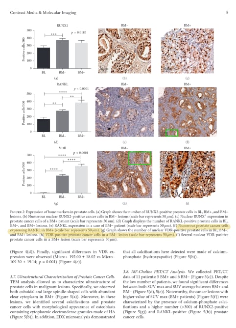
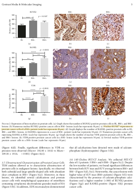
- In the article entitled “Prostate Osteoblast-Like Cells: A Reliable Prognostic Marker of Bone Metastasis in Prostate Cancer Patients” by Manuel Scimeca, Nicoletta Urbano, Rita Bonfiglio, Sarah Natalia Mapelli, Carlo Vittorio Catapano, Giuseppina Maria Carbone, Sara Ciuffa, Mario Tavolozza, Orazio Schillaci, Alessandro Mauriello and Elena Bonanno, published in the journal Contrast Media & Molecular Imaging in 2018, the authors use the same microscope image twice in Figure 2. In one case (Figure 2f) it is supposed to depict cells from patients with tumors without metastasis, while in another (Figure 2h) it is used to illustrate cells from metastatic patients.
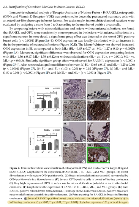
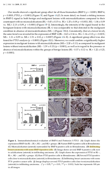
- In the article “Microcalcifications Drive Breast Cancer Occurrence and Development by Macrophage-Mediated Epithelial to Mesenchymal Transition” authored by Manuel Scimeca, Rita Bonfiglio, Erika Menichini, Loredana Albonici, Nicoletta Urbano, Maria Teresa De Caro, Alessandro Mauriello, Orazio Schillaci, Alessandra Gambacurta and Elena Bonanno, published in 2019 in the International Journal of Molecular Sciences, an image is used that supposedly depicts bone cells from breast cancer metastases.
However, the image is copied from research published in the journal Cell Death Discovery in 2018, in an article on bone cells, not authored by Schillaci and which had nothing to do with breast cancer cells. For this article, Orazio Schillaci was responsible for validating, reviewing and correcting the study.

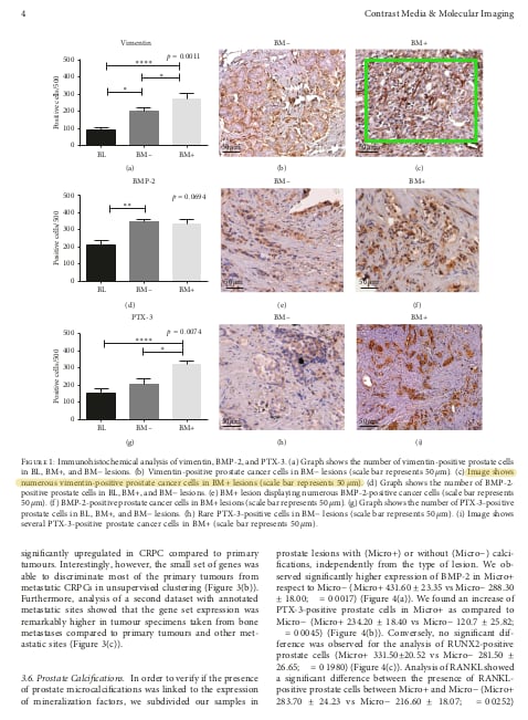
- In the article “18F-Choline PET/CT Identifies High-Grade Prostate Cancer Lesions Expressing Bone Biomarkers” by Nicoletta Urbano, Manuel Scimeca, Antonio Crocco, Alessandro Mauriello, Elena Bonanno and Orazio Schillaci, published in 2019 in the Journal of Clinical Medicine, in Figure 2 the same image is used to illustrate the presence of prostate cells positive for the BMP-2 protein and the presence of cancer cells positive for the Vitamin D receptor.
For this work, Schillaci is responsible for the supervision, conception, research, validation and first draft of the study and is listed as its “corresponding author,” i.e., the researcher to be contacted for further questions and concerns.
- In the article “Breast-Specific Gamma Imaging with [99mTc]-Sestamibi: An In Vivo Analysis for Early Identification of Breast Cancer Lesions Expressing Bone Biomarkers,” by Nicoletta Urbano, Manuel Scimeca, Carmela Di Russo, Elena Bonanno and Orazio Schillaci, published in the Journal of Clinical Medicine in 2020, an image is featured that had already been used to show the success of a completely different experiment in an article published in 2019 in the International Journal of Molecular Sciences.
Figure 2C in the 2020 article claims to show estrogen-positive tumor cells examined under an electron microscope, while the identical image in the 2019 study claims to show tumor cells positive for RANKL protein. Schillaci is listed as the corresponding author for the 2020 article, where he is responsible for supervision, conception, validation and the first draft.
- In the article “Increased 18F-choline PET/CT PET/CT Uptake in Undifferentiated Prostate Cancers with High Proliferation Index” by Nicoletta Urbano, Manuel Scimeca, Elena Bonanno, Alessandro Mauriello and Orazio Schillaci, published in Cancer Research Reports in 2020, a microscope image is featured which had already been used in a 2018 Contrast Media & Molecular Imaging article authored by Schillaci and collaborators, for which the minister is the corresponding author.
In Figure 1A of the 2020 article, the image claims to show prostate cancer cells with a high presence of two proteins (vimentin and Ki67). The same image was used in the 2018 article, claiming to show cells that were positive for a different protein, RUNX2.
- In the article “Novel Biological and Molecular Characterization in Radiopharmaceutical Preclinical Design” by Nicoletta Urbano, Manuel Scimeca, Anna Tolomeo, Vincenzo Dimiccoli, Elena Bonanno and Orazio Schillaci, published in the Journal of Clinical Medicine in 2021, a microscope image was used that is also found in a 2019 article in the International Journal of Molecular Sciences, also by Schillaci and team.
However, in the 2021 article the image supposedly shows prostate cancer cells, while in the 2019 article it claimed to show breast cancer cells. Orazio Schillaci is listed as responsible for the conception, methodology, supervision, writing, review and correction of the 2021 article.
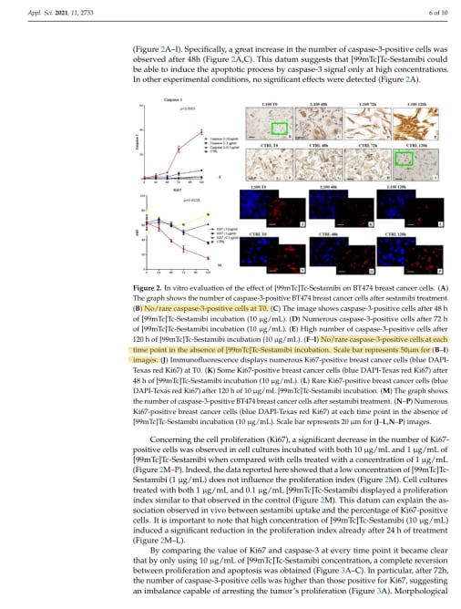
- In the article “[99mTc]-Sestamibi Bioaccumulation Can Induce Apoptosis in Breast Cancer Cells: Molecular and Clinical Perspectives” by Nicoletta Urbano, Manuel Scimeca, Rita Bonfiglio, Alessandro Mauriello, Elena Bonanno and Orazio Schillaci, published in 2021 in Applied Sciences, the same image is used twice: first claiming to show cells treated with a drug at the initial starting time of the experiment, and then, in Figure 2I, the same image, cropped differently, is used to show untreated cells used as a control at 120 hours after the start of the experiment.
For this article, Minister Schillaci is the corresponding author and was responsible for the study’s design, data, first draft and supervision.
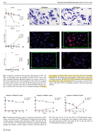
- In the article “[99Tc]Sestamibi bioaccumulation induces apoptosis in prostate cancer cells: an in vitro study” published by Nicoletta Urbano, Manuel Scimeca, Elena Bonanno, Rita Bonfiglio, Alessandro Mauriello and Orazio Schillaci in 2022 in the journal Molecular and Cellular Biochemistry, Figures 1F and 1H are the same image after a color change.
According to the captions, one electron microscope image is used to show tumor cells positive for the “caspase-3” protein after 120 hours of treatment with a drug. The other is supposed to show cells positive for another protein (“AIF”) after 24 hours of treatment. However, a computer analysis shows that the two are the same image.
Originally published at https://ilmanifesto.it/tutte-le-anomalie-delle-ricerche-schillaci on 2023-09-14